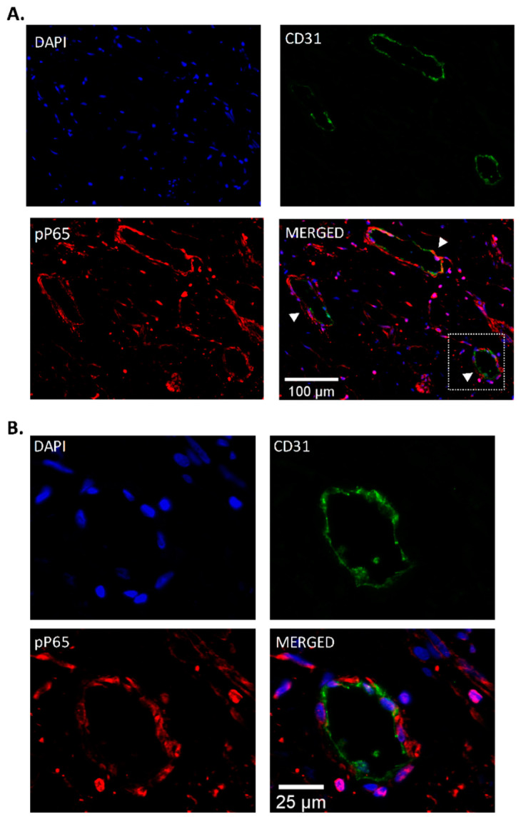
Figure 3.
Pulmonary endarterectomy (PEA) immunofluorescence. (A) Representative images showing the localization of phospho-NF-κB-P65 (pP65) in vessels in endarterectomy specimens from patients with CTEPH (n = 8), using double labeling with CD31/PECAM (green) and pP65 (red). (B) pP65 immunoreactivity was observed in endothelial cells from vessels within the thrombus (magenta, indicated by the white arrows). Nuclei were counterstained with DAPI (blue). Scale bar, 100 μm (panel (A)) and 25 μm (panel (B)).

