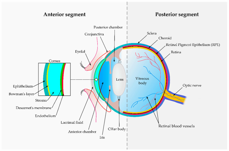
Figure 1.
Schematic representation of the ocular anatomy. An imaginary line (dotted grey vertical line) divides the ocular structures into two segments. The anterior segment contains the cornea (detailed within the insert), iris, ciliary body and both anterior and posterior chambers. The aqueous humour fills the anterior and posterior chambers, while most of the posterior segment is filled by a hyaluronan-rich gel known as vitreous humour. The posterior segment also contains the sclera, similar in composition to the corneal stroma. The choroid is a highly vascularised tissue separated from the retina (neural tissue) by a monolayer of hexagonal pigment-containing cells: the retinal pigment epithelium.

