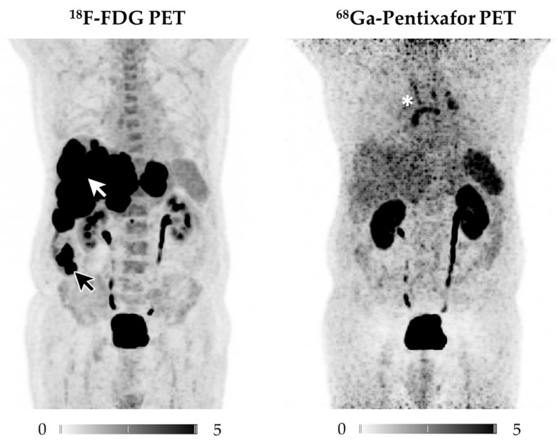
Figure 1.
Displayed are Maximum Intensity Projections (MIP) of the 18F-FDG (left) and 68Ga-Pentixafor PET scans of patient #2. Whereas 18F-FDG depicts the ileal primary (black arrow) as well as multiple liver metastases (white arrow), none of the tumor manifestations are revealed by CXCR4-directed PET imaging. Incidental finding: The mediastinal tracer uptake in 68Ga-Pentixafor PET (white star) was traceable to enlarged mediastinal lymph nodes, most likely due to chronic lung fibrosis and not related to NEC, as follow-up imaging confirmed.

