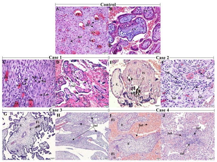
Figure 1.
Histopathological analysis of the aborted materials detected an inflammatory environment: (A) abortion material from a healthy patient without any infectious disease history during pregnancy (control) showing decidual cells (DC) and (B) decidual vessels (DV) with the regular aspects. (C) Case 1 exhibiting decidua with mononuclear and polymorphonuclear—lymphocytes (Ly) and neutrophils (Nø) (D) and decidua with inflammatory infiltrate (Inf). (E) Case 2 with villous edema (E), the deposition of fibrinoid material (Fi) and calcification (C) (F) and decidua with intense inflammatory infiltrate. (G) Case 3 with dysmorphic villi with edema, focal areas of the deposition of fibrinoid material and calcification (H) and deciduitis. (I) Case 4 with intervillous space with inflammatory cells, villous edema and areas of the deposition of fibrinoid (J) and deciduitis. 10 µm = 1000×, 20 µm = 400×, 50 µm= 200×, 100 µm = 100×.

