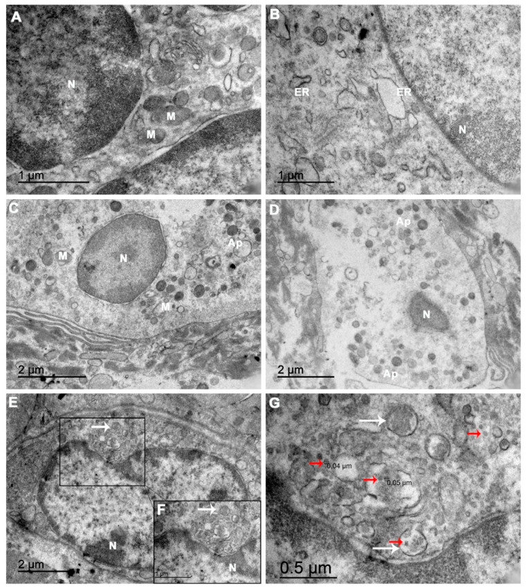
Figure 4.
An electron microscopy analysis of the ultrathin abortion material sections showed damaged organelles and virus-like particles: (A) Electron micrographs of CHIKV-infected cells showing dispersed chromatin in the nuclei (N), mitochondria (M) with fewer cristae and (B) endoplasmic reticulum (ER) exhibiting dilated cisterns. (C) A decidual cell presenting a rarefied cytoplasm with an absence of organelles and starting to produce apoptotic bodies (Ap). (D) A decidual cell in apoptosis. (E,F) A cell with vesicles surrounding a cluster of virions in the cytoplasm (white arrows). (G) In the same field, at a higher magnitude, these CHIKV virus-like particles (red arrows) are located near a ruptured ER. The scale bar indicates that the particles are approximately 40–50 nm in size, consistent with the CHIKV.

