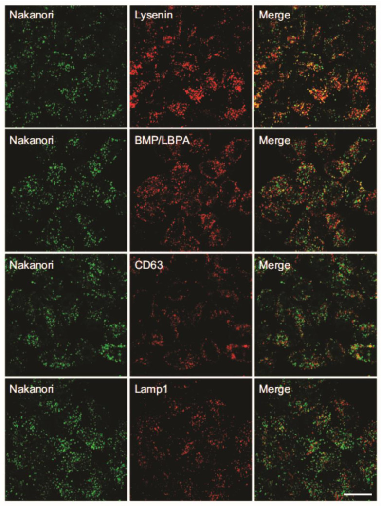
Figure 4.
Intracellular distribution of nakanori and organelle markers. Representative fluorescent images of double immunolabeling of Hela cells with nakanori (green) and organelle markers (red) showing co-localization of nakanori with lysenin, and with markers of the internal membranes of late endosomes, BMP/LBPA (bis(monoacylglycero)phosphate/lysobisphosphatidic acid) and CD63. Some co-localization of nakanori was seen with Lamp 1, as a marker of the limiting membrane of late endosomes. Scale bar: 20 μm. Adapted from [40].

