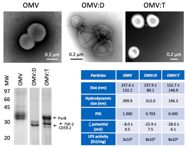
Figure 1.
Characterization of Outer Membrane Vesicles (OMV) and OMV–antigen complexes. TEM images of unconjugated biotinylated OMV of N. lactamica (OMV, upper left), OMV conjugated with rRzvSmCD59.2 (OMV:D, upper center) and OMV conjugated with rRzvSmTSP-2 (OMV:T, upper right). Lower left panel: electrophoretic mobility of OMV and OMV–antigen complexes. Left arrows indicate the position of the main N. lactamica protein PorB, of SmTSP-2 in OMV:T and of SmCD59.2 in OMV:D (the two antigens having a calculated MW of 27.2 kDa for rRzvSmTSP-2 and 26.9 kDa for rRzvSmCD59.2). Lower right table: Summary of the OMV characteristics of size (measured in TEM, mean ± SD of 16–131 particles), hydrodynamic size, polydispersity, ζ-potential (mean of 3 determinations ± SD) (all measured by dynamic light scattering (DLS)) and LPS activity (measured with the LAL assay). PDI, polydispersity index.

