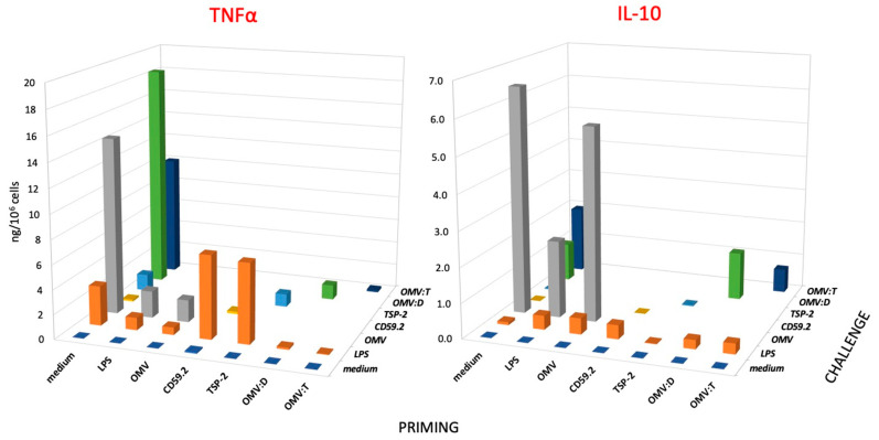
Figure 3.
Secondary response of human monocytes to challenge with S. mansoni antigens. Production of TNFα (left panel) and IL-10 (right panel) of human monocytes that had been previously exposed (PRIMING in the horizontal axis) to culture medium alone (medium), LPS (1 ng/mL), unconjugated OMV (OMV), rSmCD59.2 (CD59.2), rSmTSP-2 (TSP-2), OMV:D, or OMV:T (all at 0.1 μg antigen/mL). After 6 days of resting, cells were challenged (see depth axis CHALLENGE) with a 10x higher concentration of stimuli; medium (purple), LPS (orange), OMV (gray), rSmCD59.2 (yellow), rSmTSP-2 (light blue), OMV:D (green) and OMV:T (dark blue). LPS was used as control challenge for cells primed with every kind of stimuli. Data are the values of the 24-h cytokine production by cells from one donor representative of three examined (see Supplementary Table S3 for the values of individual donors). SD of technical replicates were always <10% and are not shown. Statistical significance is reported in the Supplementary Table S4.

