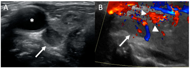
Figure 2.
Ultrasound: 54-year-old woman with primary hyperparathyroidism (PHPT). Transverse grayscale (A) ultrasound image demonstrates an ovoid hypoechoic lesion (white arrow) posterior to the right lobe of the thyroid gland and carotid artery (*) consistent with a parathyroid adenoma. (B) Color doppler ultrasound image shows a polar feeding vessel with peripheral vascularity (white arrowheads).

