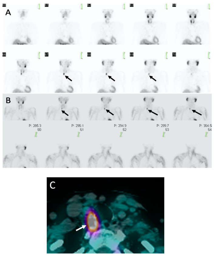
Figure 3.
Sestamibi: 62-year-old woman with PHPT. (A) Coronal early phase sestamibi single photon emission computed tomography (SPECT) images demonstrate uptake in both the thyroid and parathyroid glands with asymmetric contour along the posterior right thyroid gland (black arrows).(B) Coronal delayed phase sestamibi SPECT images demonstrate radiotracer washout from the thyroid gland with retention of radiotracer posterior to the right thyroid gland consistent with a right upper parathyroid adenoma (black arrows) (C) Axial delayed phase sestamibi SPECT/computed tomography (CT) shows 99mTechnetium-sestamibi (MIBI) uptake posterior to right thyroid gland (white arrow) corresponding with a right upper parathyroid adenoma. Addition of CT to SPECT allows for improved anatomic localization.

