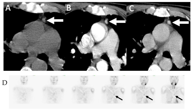
Figure 5.
Ectopic mediastinal adenoma: 62-year-old man with primary hyperparathyroidism 4D-CT demonstrating a small enhancing nodule in the anterior mediastinum (arrows) seen on non-contrast (A), arterial-phase (B) and delayed phase (C) imaging without washout. (D) Coronal early phase sestamibi SPECT images demonstrate mild uptake in the anterior mediastinum, corresponding to the enhancing nodule on 4D-CT. The patient was subsequently taken for thoracoscopic resection which demonstrated an ectopic parathyroid adenoma within thymic tissue.

