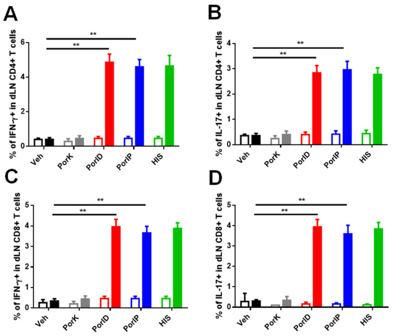
Figure A3.
Percentage of functional CD4+ and CD8+ T cells in skin dLN. C57BL/6J mice were injected intradermally in the ear with vehicle (Veh), proteinase K-digested porins (PorK), porins (PorID), or intraperitoneally with porins (PorIP) or heat-inactivated S. Typhi (HIS) at day 0. All groups were boosted at day 14 with porins injected intradermally in the ear. Count of the percentage of IFN-γ- (A) and IL-17- (B) producing CD4+ T cells found in skin dLN at day 28. Same populations percentages were analyzed for skin dLN CD8+ T cells (C,D) (n = 7; 2 independent experiments; significant difference for a Kruskal–Wallis test ** p < 0.05 * p < 0.1 with Dunn’s multiple comparison).

