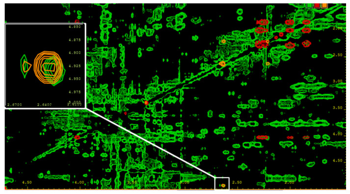
Figure 4.
2D TOCSY of a serum sample from an AML patient with an IDH2-R140Q mutation. For the removal of macromolecules, the serum was filtered using a Nanosep filtering device with a 3 kDa cutoff. Signal assignment was based on comparison with reference spectra of pure compounds. Green = serum sample; red = 2-HG reference spectrum; and orange = 2-HG-lactone reference spectrum. Besides overlapping signals for both compounds, unique signals are present, as shown in the white box for the 2-HG-lactone, and allow for the discrimination of the two metabolites. In NMR spectra of probands carrying no IDH1/2 mutation, neither 2-HG nor its lactone could be detected.

