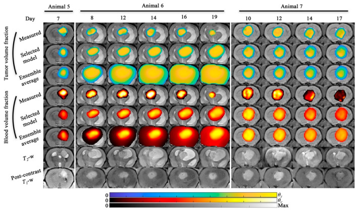
Figure 7.
Model predictions (scenario 2) for animal 5, 6, and 7. Model predictions of tumor and blood volume fractions at the central slice are shown for animal 5, 6, and 7. For all animals, the model predictions from the selected model and the ensemble average are shown. The bottom two rows show the T2-weighted and post-contrast T1-weighted MRI. For animal 5, only one prediction time point was available and both the selected and ensemble average model resulted in <8.7% error in tumor volume predictions. For animal 6, tumor growth was predicted from day 8 to 19 and resulted in 16.0% and 521% error in tumor volume predictions for the selected and ensemble models, respectively. For animal 7, four prediction time points were available and both the selected and ensemble average model resulted in <21.5% in tumor volume predictions across all time points. A sagittal view of this figure is shown in Figure S2.

