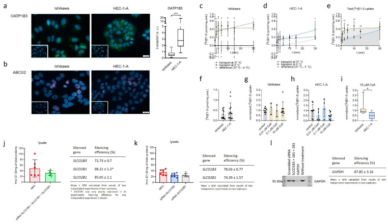
Figure 2.
Immunocytochemical staining of OATP1B3 and ABCG2 and uptake of E1-S in Ishikawa and HEC-1-A. (a) Levels of OATP1B3 immunoreactivity in EC cell lines. Mann–Whitney U test after evaluation of 30 randomly chosen fields from three independent experiments (10 fields from each experiment). (b) Levels of ABCG2 immunoreactivity in EC cell lines. Representative pictures of ABCG2 staining are shown. Insets show corresponding cell lines incubated with normal rabbit (a) or mouse (b) serum. (c,d) Time-course of 16 nM [3H]E1-S uptake at 37 °C (total transport) and 4 °C (diffusion), measured in cell lysates in three individual experiments in duplicates. Transporter-mediated uptake of E1-S was calculated by subtraction of average values at individual temperatures. (e) Comparison of total E1-S uptake in Ishikawa and HEC-1-A over 30-min time-course. (f) Absolute uptake of E1-S after 30 min (Mann–Whitney U Test). (g,h) Normalized uptake of 16 nM [3H]E1-S after 30 min incubation with different concentrations of OATP inhibitor cyclosporine A (CsA). (i) E1-S uptake in the presence of 10 µM CsA. (Mann–Whitney U test). (j) Uptake of [3H]E1-S (2 min) after 48-h transient transfection with small-interfering RNAs targeting combination of genes SLCO1B3, SLCO1B1 and SLCO2B1 in HEC-1-A cell line (results of two independent experiments in three replicates (Mann–Whitney U test). (k) Effect of silencing of individual genes, SLCO1B3 or SLCO1B1 on 2 min [3H]E1-S uptake evaluated after 72-h siRNA transfection (two independent experiment in three replicates, Kruskal–Wallis with Dunn’s Multiple comparisons). (l) Western blot experiment confirming silencing of a control GAPDH (silencing efficiency (%) mean ± SEM, 87.85 ± 3.16) in two independent experiments in three replicates. In all experiments, silencing efficiency was calculated based on the expression of evaluated genes in NEG, cells treated with scrambled siRNA. Results of E1-S uptake were normalized to the total amount of proteins in cell lysates in individual treatments. Data are shown as mean ± SD. ir-area—immunoreactive area; a. u.—arbitrary unit, ns—p-value > 0.05, * p-value ≤ 0.05, ** p-value ≤ 0.01. *** p-value ≤ 0.001.

