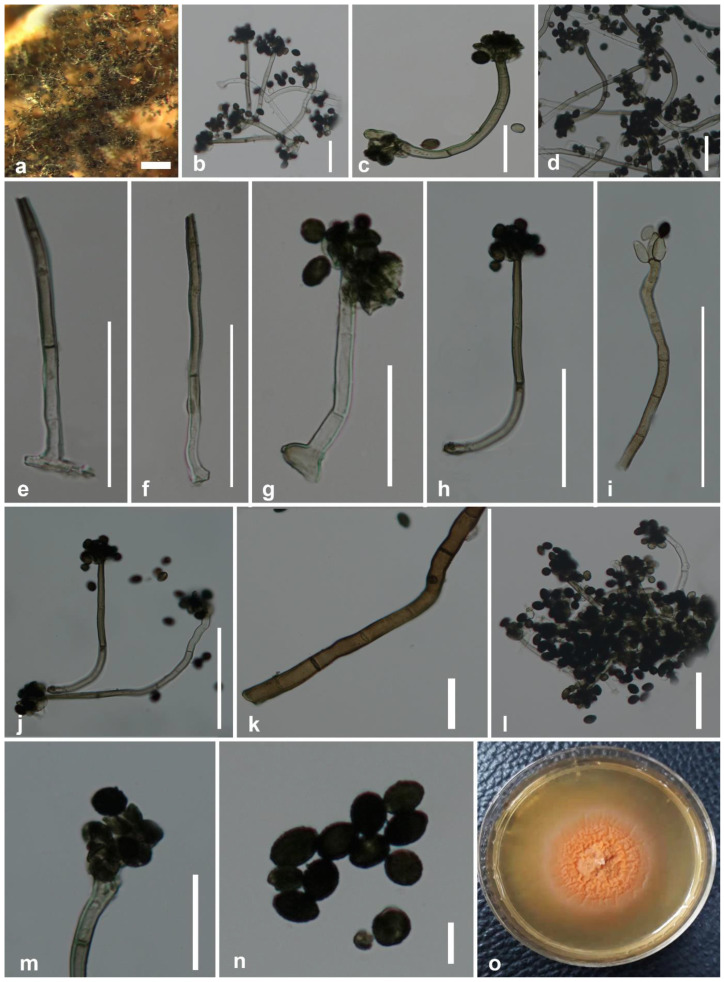
Figure 2.
Stachybotrys musae (MFLU 20–0626, holotype). (a) Conidiophores on the substrate surface; (b–d,g,h,j,m) conidiophores with attached conidia; (e,f) conidiophores; (i) conidiophore with monophialidic conidiogenous cells; (k) mycelium; (l) mass of conidia and conidiophores; (n) conidia; (o) colonies on PDA after 8 weeks. Scale bars: (a) = 500 μm; (j) = 200 μm; (c–i,l,m) = 50 μm; (b,f,g) = 25 μm; (n,k) = 5 μm.

