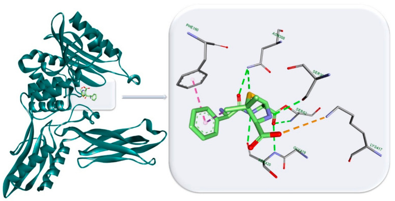
Figure 3.
Penicillin binding protein 4 (PBP4) from E. coli in complex with ampicillin (light green): HBs (Ser 30, Ser 62, Ser 420 and Asn 308) are depicted as green dotted lines, Pi-hydrophobic interactions (Phe 160) are depicted as purple dotted lines and electrostatic interactions (Lys 417) as orange dotted lines. The figure was built with Biovia Discovery Studio 4.1 using the PDB entry 2EX6 [29].

