
Figure 2.
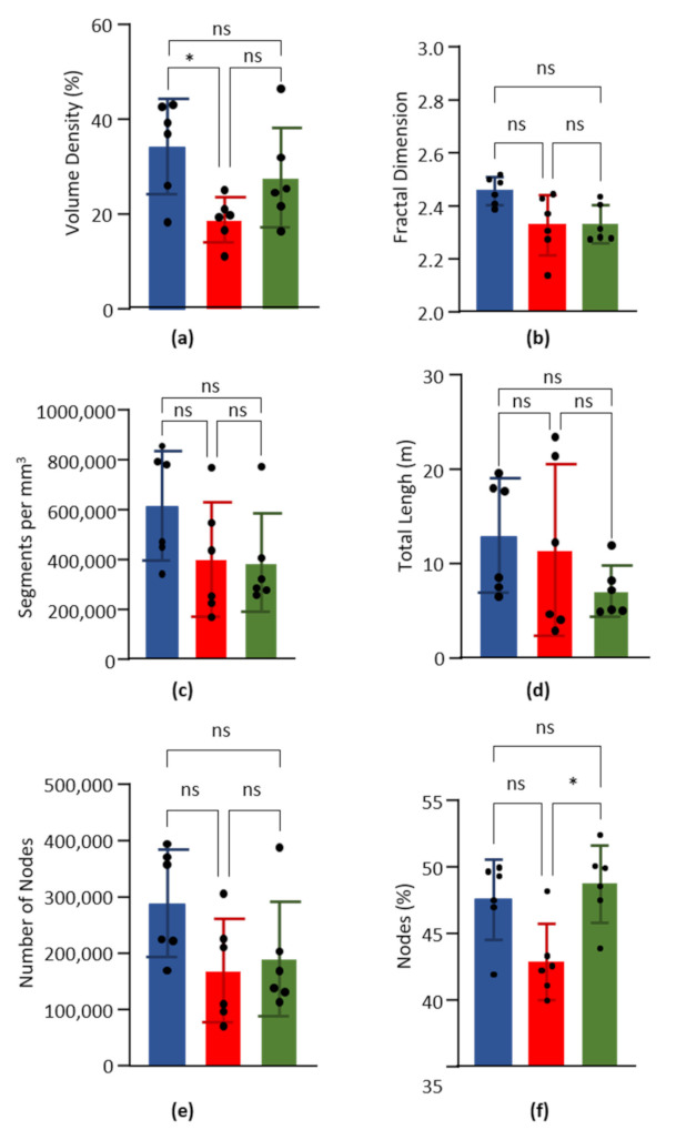
Global parameters. Data obtained from the left ventricle (blue), septum (red) and right ventricle (green) of six hearts. Columns are means and error bars are standard deviations. Black dots are individual values. Data were compared by Kruskal–Wallis test with post-hoc Dunn’s test. ns = non significant. * p < 0.05. (a). Volume capillary density, in %. (b). Fractal dimension. (c). Number of capillary segments per mm3 of cardiac tissue. (d). Total length of capillary segments (m) per mm3 of cardiac tissue. (e). Number of nodes per mm3 of cardiac tissue. (f). Percentage of nodes on the number of segments.