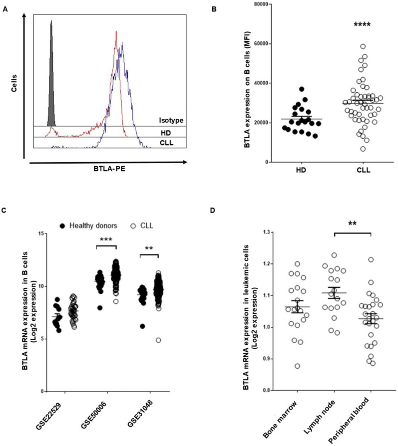
Figure 1.
BTLA immune checkpoint expression is upregulated on leukemic cells from patients with CLL. BTLA surface expression on leukemic cells from patients with CLL (n = 46) and B cells from HD (n = 20) was evaluated by flow cytometry. (A) Representative histogram of BTLA expression (MFI) in HD and patients. (B) Comparison of BTLA levels between leukemic cells from patients and B cells from controls (MFI ± SEM). (C) Three available microarray data from the GEO database were interrogated to analyze BTLA mRNA expression in leukemic cells from patients with CLL in comparison with HD. (D) BTLA mRNA levels were evaluated according to leukemic cell localization (GSE21029). Each dot represents an individual sample. ** p < 0.01, *** p < 0.001 and **** p < 0.0001.

