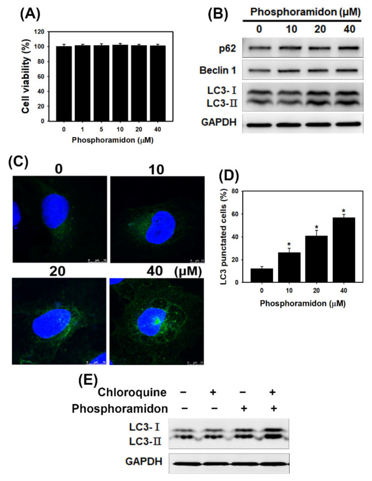
Figure 3.
Phosphoramidon triggers autophagy in HK-2 cells. (A) Cell viability of phosphoramidon-treated cells. Cells were treated with several concentrations of phosphoramidon for 24 h. Data were presented as the means ± standard deviation of three independent experiments. (B) The protein levels of p62, beclin 1 and LC3 in HK-2 cells treated with phosphoramidon for 24 h. (C) Imaging of LC3 by confocal immunofluorescence microscopy following 24 h of treatment with phosphoramidon. Scale bar = 10 µm. (D) Quantification of punctate LC3 staining. Cells were treated with various concentrations of phosphoramidon for 24 h. * p < 0.05 compared with the control. Data were presented as the means ± standard deviation of three independent experiments. (E) Western blotting of LC3-I and LC3-II expression in HK-2 cells. The cells were pretreated with chloroquine (5 µM) for 1 h and then treated with phosphoramidon (20 µM) for 24 h.

