Figure 2.

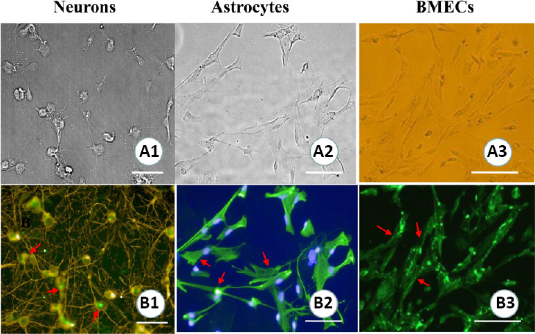
Cell morphological characteristics and the identification of a hippocampal neurovascular unit.
(A1–3) Cell morphologies of hippocampal neurons, astrocytes, and brain microvascular endothelial cells (BMECs). (B1–3) The identification of neurons, astrocytes, and BMECs (immunofluorescence staining). Arrows show the positive expression of neuron-specific enolase (B1), glial fibrillary acidic protein (B2), and platelet endothelial cell adhesion molecule-1/CD31 (B3). Scale bars: 100 µm.
