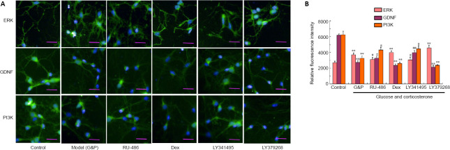
Figure 6.
Aberrant immunopositivity against ERK, BDNF, and PI3K in hippocampal neurovascular unit neurons after diabetes-related depression induction.
(A) Typical images showing ERK, BDNF, and PI3K immunopositivity (immunofluorescence staining). The detection of ERK, BDNF, and PI3K is shown in green, stained by fluorescein isothiocyanate. Simulated DD conditions (G&P) significantly upregulated ERK and down-regulated GDNF and PI3K when compared with the control conditions. The upregulation of ERK and the downregulation of GDNF and PI3K could be reversed by both RU-486 and LY341495 treatment when compared with the G&P group. However, dexamethasone and LY379268 aggravated the disordered expression patterns of these proteins. Scale bars: 100 µm. (B) Quantitative analysis of ERK, BDNF, and PI3K immunopositivity. Data are expressed as the mean ± SD (n = 3). **P < 0.01, vs. control group; #P < 0.05, ##P < 0.01, vs. model group (G&P) (one-way analysis of variance followed by Dunnett’s post hoc test). Experiments were performed in triplicate. BDNF: Brain-derived neurotrophic factor; Dex: dexamethasone, i.e., glucocorticoid receptor agonist; ERK: extracellular signal-regulated kinase; G&P: glucose and corticosterone, i.e., simulated diabetes-related depression conditions; LY341495: metabotropic glutamate receptor 2/3 receptor blocker; LY379268: metabotropic glutamate receptor 2/3 receptor agonist; PI3K: phosphoinositide 3-kinase; RU-486: Mifepristone, i.e., glucocorticoid receptor blocker.

