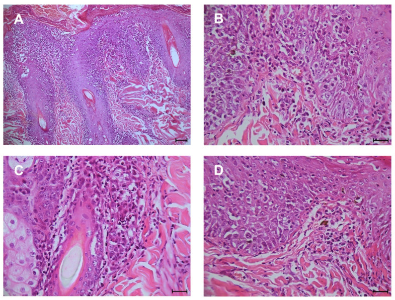
Figure 1.
Histopathologic features of DLE. (A) interface dermatitis (bar 75 µm); (B,C) infiltrate of plasma cells and lymphocytes within dermo-epidermal junction and hydropic degeneration of keratinocytes of stratum basale (scale bar 25 µm); (D) scattered macrophages contain melanin (pigmentary incontinence) within the superficial dermis (scale bar 25 µm).

