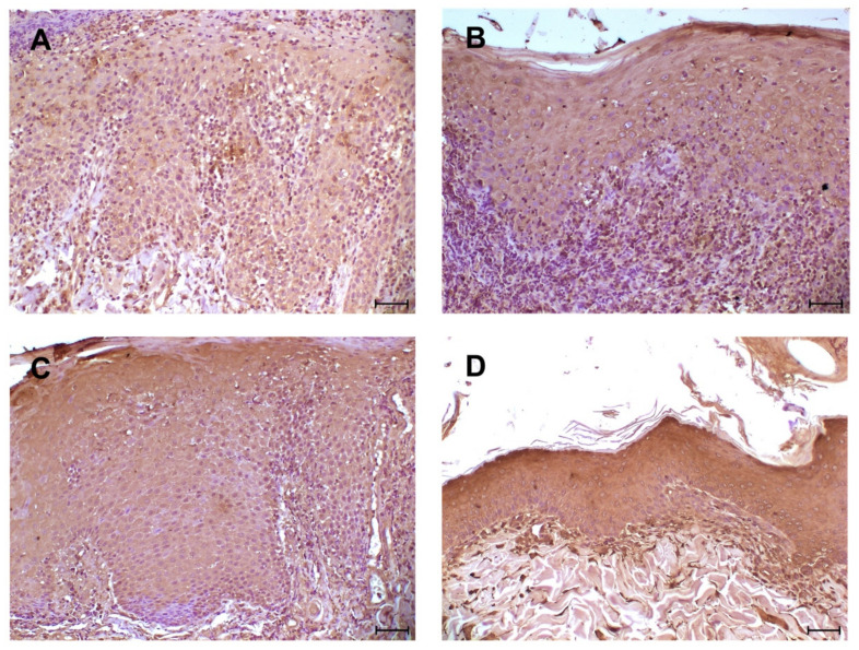
Figure 3.
Immunohistochemical analysis for TLR4: four different cases of DLE with epidermis diffusely positive; (A) moderate intensity staining of the immunolabeled; (B–D) high-intensity staining of inflammatory cells. Diffuse immunolabelling of inflammatory cells at dermo-epidermal junction. (A–D) scale bar 50 µm.

