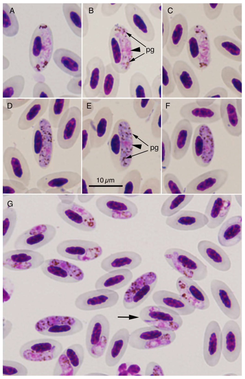
Figure 1.
Haemoproteus columbae gametocytes in the blood of farmed domestic pigeons (Columba livia f. domestica) from Yogyakarta Special Region, Indonesia. Mature microgametocytes (A–C), mature macrogametocytes (D–F), and various developmental stages of gametocytes (G) in erythrocytes of a pigeon from Ngemplak, Yogyakarta. All photographs are at the same magnification, and the scale bar (10 µm) is shown in the photograph (E). Nuclei (arrowheads) and pigment granules (pg) of H. columbae are shown in the photographs (B,E). Arrow in the photograph (G) indicates an erythrocyte with two gametocytes.

