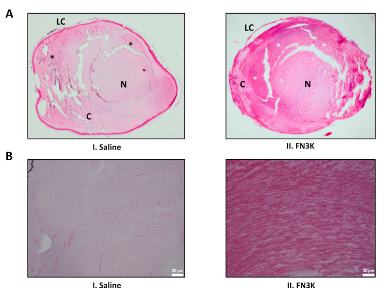
Figure 6.
(A) Hematoxylin and eosin-stained sections of a saline (I) and FN3K treated (II) lens originating from the same ob/ob mouse. Disorganized and organized zones of lens fibers are indicated with black and white asterisks, respectively. C, cortex; LC, lens capsule; N, nucleus. (B) Close-up on the cortical lens fibers of the saline (I) and FN3K treated (II) lens.

