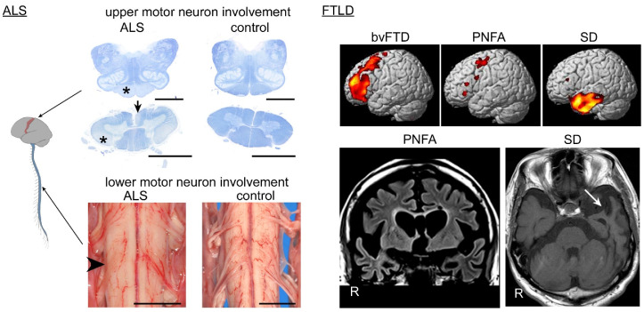
Figure 1.
Systemic atrophy of central nervous system in amyotrophic lateral sclerosis (ALS) and frontotemporal lobar degeneration (FTLD) patients. The spinal cord and medulla oblongata are stained with Klüver–Barrera method. Involvement of the upper motor neurons results in tract degeneration of the pyramidal tract in the medullary pyramid and lateral column (*) and anterior cerebrospinal fasciculus (arrow) of the spinal cord; the change is usually prominent in the caudal segments of the spinal cord. Involvement of the lower motor neurons results in atrophy of the anterior roots in the spinal cord (arrowhead); the anterior roots are thin and hardly visible, compared with the dorsal roots. Scale bars = 5 mm. Cerebral MRI illustrates a vulnerable region corresponding to each clinical subtype: the prefrontal area for behavioral variant frontotemporal dementia (bvFTD), the para-Sylvian operculum and primary motor cortex for progressive non-fluent aphasia (PNFA), and the anterior portion of the unilateral (dominant hemisphere) temporal lobe for SD (arrow).

