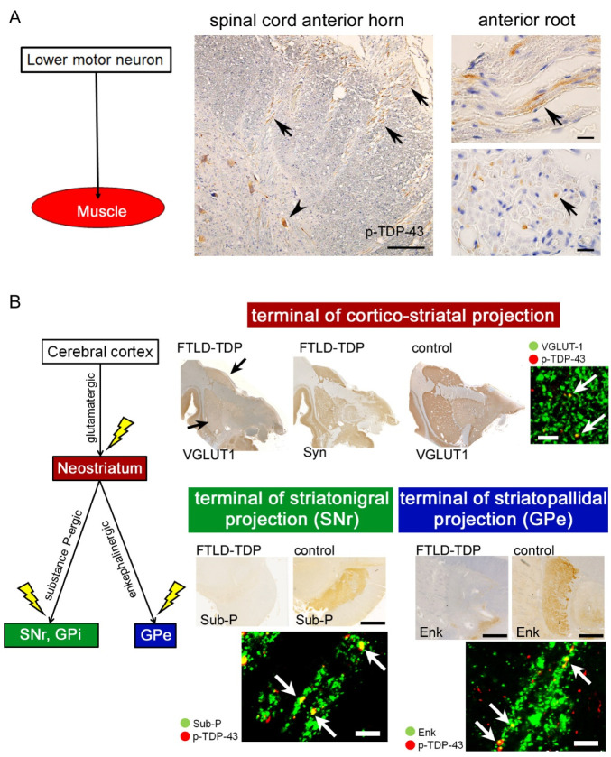
Figure 3.
TDP-43 pathology in multi-system axons and axon terminals. The upper section (A) demonstrates the spinal cord of an ALS-TDP patient who died six months after the disease onset. Phosphorylated TDP-43 (p-TDP-43) aggregated not only in the anterior horn neurons (arrowhead) but also in the anterior roots (arrows). Scale bars: 100 μm for the panel of the anterior horn and 10 μm for the panels of the anterior roots. The lower section (B) displays pathologic changes of the cortico-striatal circuit in FTLD-TDP patients. Axon terminals of the corticofugal neurons were visualized with anti-VGLUT-1 immunohistochemistry (IHC) in the neostriatum. Those of the striatofugal neurons were labeled with anti-enkephalin (Enk) IHC in the external segments of the globus pallidus (GPe) or with anti-substance-P (Sub-P) IHC in the internal segment of GP (GPi) and pars reticulata of the substantia nigra (SNr). Patients with FTLD-TDP displayed loss of those axon terminals and p-TDP-43 aggregation within the pre-synaptic buttons. Comparing the loss of VGLUT-1-immunopositive terminals in the neostriatum (arrows) and sparing of synaptophysin (Syn) immunostaining indicates specific loss of cortico-striatal projections but spares of other projections. Scale bars: 10 μm.

