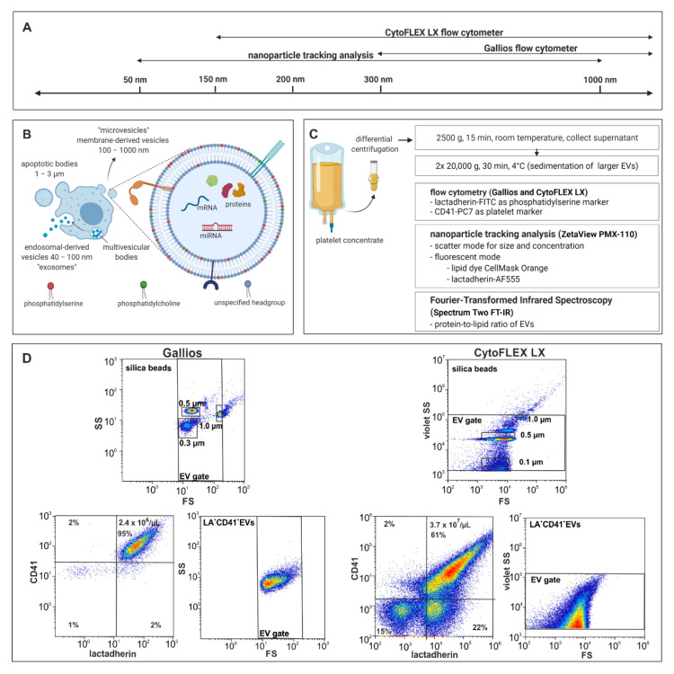
Figure 1.
Characterization of EVs by flow cytometry and NTA. (A,B) Size-dependent resolution limits of the devices and approximate size range of EV subpopulations (exosomes, microvesicles, and apoptotic bodies). (C) Enrichment and characterization of platelet-derived EVs. Platelet-derived EVs were enriched from medical grade platelet concentrates by differential centrifugation as described in the Methods section and characterized by flow cytometry using lactadherin as a marker of phosphatidylserine expressing EVs and CD41 as platelet marker. Further analysis was performed by NTA in scatter mode and in fluorescence mode after staining of EVs with CellMask™ Orange (CMO) and lactadherin-Alexa Fluor™ 555 (LA-AF555). The protein-to-lipid ratio was assessed by Fourier-transformed infrared spectroscopy. (D) Representative images of EV gating and scatter plots for the CytoFLEX LX vs. Gallios flow cytometers. Phosphatidylserine exposing platelet-derived EVs were identified as lactadherin+ and CD41+ events in the EV gate. Figure 1A–C was created with BioRender.com (accessed on 5 April 2021).

