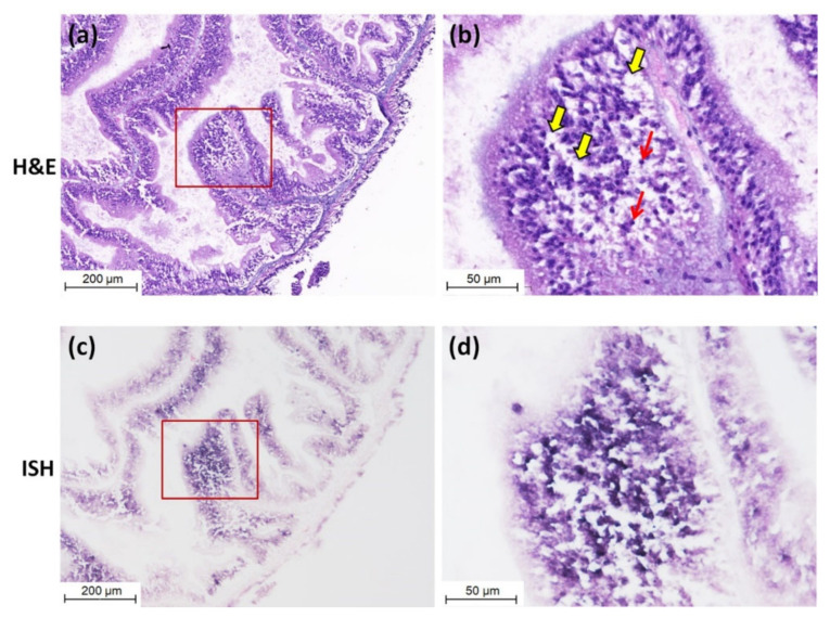
Figure 3.
Micrographs of hematoxylin and eosin-phloxine (H&E) staining and in situ hybridization (ISH) of the intestine of sea cucumber naturally infected with CMNV. (a) Micrographs of H&E staining of the intestine. (b) Magnified micrographs from the red-framed areas of (a). Karyopyknosis (red arrows) and extensive vacuolation (yellow arrows) were observed in the intestinal epithelial cells. (c) Micrographs of ISH of the intestine. (d) Magnified micrographs from the red-framed areas of (c). Intense CMNV positive hybridization signals (colored deep-purple) were detected in the intestinal epithelial cells. Scale bars: (a,c) 200 µm, (b,d) 50 µm.

