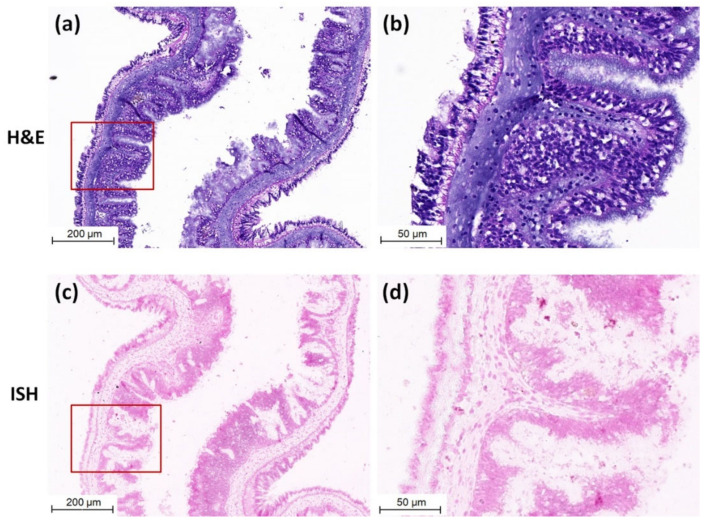
Figure 4.
Micrographs of H&E staining and ISH of the intestinal tissue of sea cucumber of negative control. (a) Micrographs of H&E staining of negative control intestinal tissue. (b) Magnified micrographs from the red-framed areas of (a). No obvious histopathological change was observed in the intestinal tissue. (c) Micrographs of ISH of negative control intestinal tissue. (d) Magnified micrographs from the red-framed areas of (c). No CMNV positive hybridization signal was detected in the intestinal tissue. Scale bars: (a,c) 200 µm, (b,d) 50 µm.

