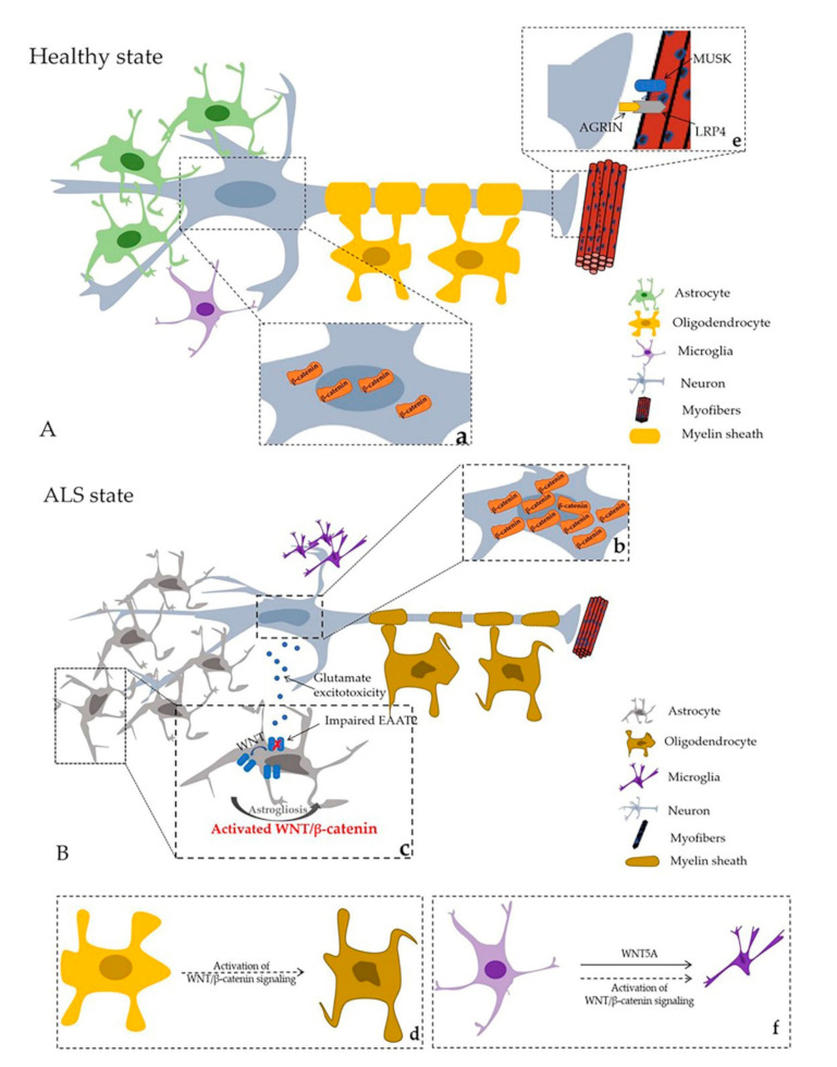
Figure 2.

Healthy state and ALS state in central nervous system (CNS) and neuromuscular junction (NMJ). (A) Healthy state; (B) ALS state; a. and b represent β–catenin distribution in healthy and ALS states, respectively; a. Normal β–catenin distribution in motor neurons. b. Extensive β–catenin accumulates in the cytoplasm in the motor neuron cell model in vitro. β–catenin protein nucleus translocation can be observed in the later stage of ALS in SOD1–G93A mice. c. Astrogliosis and molecular and functional abnormalities of astrocytes in ALS. d. Axon damage and oligodendrocyte degeneration. e. AGRIN–LRP4–MUSK complex promote stability of postsynaptic differentiation. f. WNT signaling and proinflammatory of microglia (black arrows represent phenomena in ALS; dashed arrows illustrate hypothetical modes in ALS).
