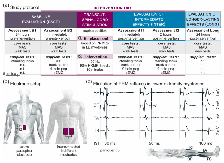
Figure 1.
Overview of the methodology. (a) Study protocol comprising a baseline evaluation (Base) with two assessments, one conducted ~24 h (B1) and the other one immediately (B2) before a 30-min session of 50-Hz transcutaneous (transcut.) spinal cord stimulation; an evaluation of intermediate carry-over effects (Inter) with two assessments, one conducted immediately (I1) and the other one two hours (I2) post-intervention; and an evaluation of longer-lasting carry-over effects (Long) of the stimulation conducted ~24 h post-intervention. Each assessment included, as core tests, the clinical determination of lower-extremity muscle hypertonia based on the Modified Ashworth Scale (MAS) and, in the ambulatory participants, the timed 10-m walk test, the timed up-and-go test, and the 2-min walk test. Supplementary (supplem.) tests were conducted as indicated in B1 or B2 as well as in I1 and I2, and included standing tasks, the trunk control test, the timed nine-hole-peg test, and a surface-EMG (sEMG) based assessment of lower-extremity spasticity. Transcutaneous spinal cord stimulation was applied with the participants lying supine. First, effective electrode (el.) placement (cf. (b)) over the lumbosacral spinal cord was confirmed based on the elicitation of posterior root-muscle reflexes (PRMRs; cf. (c)) in lower-extremity myotomes. Second, for the intervention, transcutaneous spinal cord stimulation was applied for 30 min at 50 Hz and with an amplitude corresponding to 90% of the lowest PRMR threshold. n.t., not tested. (b) Electrode setup with the active paraspinal electrode placed longitudinally over the spine, covering T11 and T12 spinal processes, and a pair of interconnected indifferent electrodes on the lower abdomen. (c) Verification of effective stimulation site over the lumbosacral spinal cord by the elicitation of PRMRs in rectus femoris (RF), biceps femoris (BF), tibialis anterior (TA), and triceps surae (TS), which demonstrate a characteristic suppression at interstimulus-intervals (ISIs) of 30 ms and 50 ms when tested by paired pulses [39]. At interstimulus-intervals of 100 ms, the responses to the second stimulus had partially recovered. Shaded backgrounds mark times of stimulus application. Three repetitions each superimposed; participant 5.

