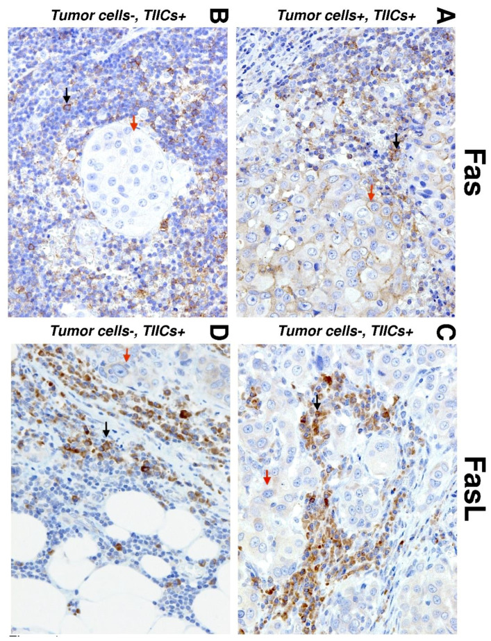
Figure 1.
Immunohistochemistry (IHC) of Fas and Fas ligand (FasL) expression in salivary gland carcinoma (SGC) tissues. Representative images show Fas (left) and FasL (right) staining in tumor cells (red arrows) and tumor-infiltrating immune cells (TIICs) (black arrows) in the tumor periphery. (A) IHC shows positive Fas staining in TIICs and tumor cells (20×). (B) IHC shows negative Fas staining in tumor cells and positive Fas staining in TIICs (20×). (C) IHC shows positive FasL staining in tumor cells and TIICs (20×). (D) IHC shows negative FasL staining in tumor cells and positive FasL staining in TIICs (20×).

