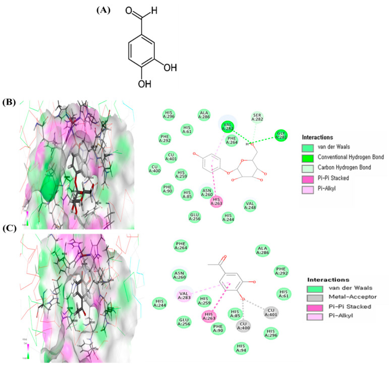
Figure 1.
Chemical structure of protocatechuic aldehyde (PA) (A) and the specific interactions between PA and tyrosinase after automated docking of PA to the tyrosinase enzyme binding site. Predicted 3D structure of the tyrosinase (Protein Data Bank; PDB code: 2Y9X)–arbutin complex and 2D diagram (B). Predicted 3D structure of the tyrosinase (PDB 2Y9X)–PA complex and 2D diagram (C). Binding energy values were obtained from the Discovery Studio (DS) 3.0 binding energy calculation program.

