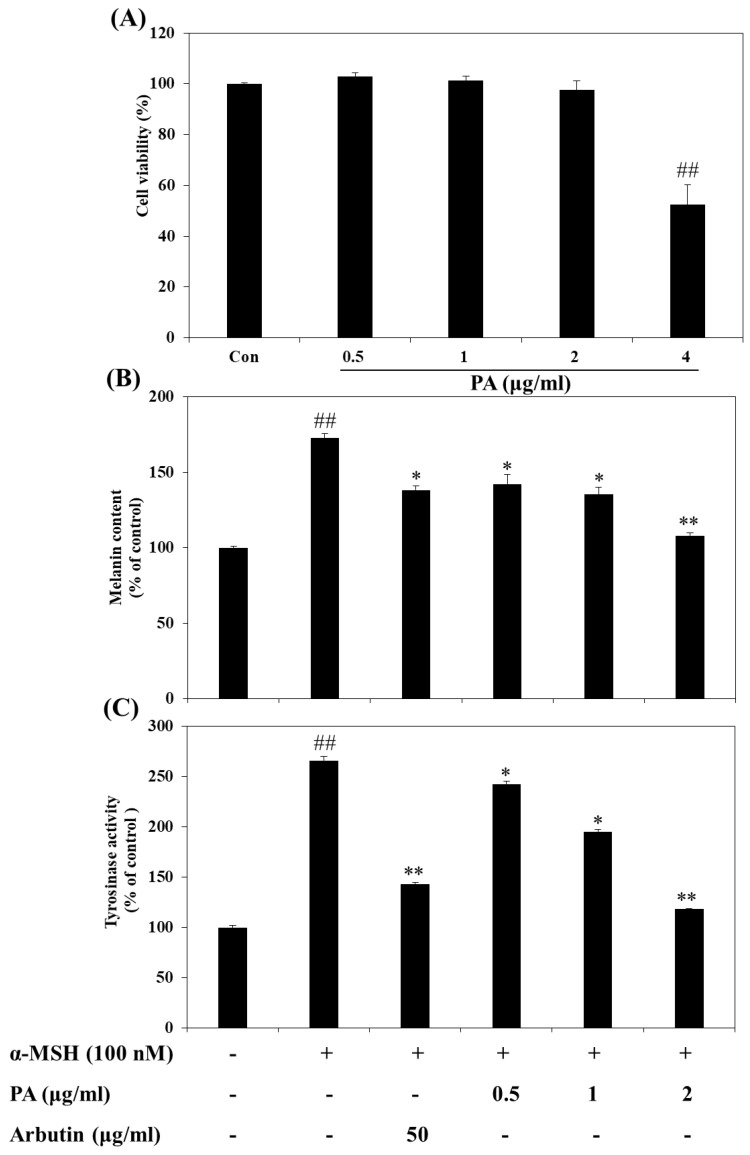
Figure 2.
Effects of PA on cellular melanin synthesis and intracellular tyrosinase activity in α-melanocyte stimulating hormone (α-MSH)-stimulated B16F10 cells. Cells were treated with the indicated concentrations of PA for 72 h and then cell viability was determined by the 3-(4,5-dimethylthiazol-2-yl)-2,5-diphenyltetrazolium bromide (MTT) assay (A). The relative cellular melanin content (B) and intracellular tyrosinase activity (C) were measured at 72 h after treatment. Cells were exposed to 100 nM α-MSH in the presence of PA at the indicated concentrations or 50 µg/mL arbutin. Values are expressed as means ± SDs of triplicate experiments (n = 3). Note: ## p < 0.01 compared to the untreated control group; * p < 0.05 and ** p < 0.01 compared to the α-MSH only group.

