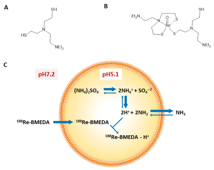
Figure 2.
Chemical structure of BMEDA (A) and 188Re-BMEDA (B). Diagram (C) depicting after-loading method for liposomes containing ammonium sulfate pH gradient radiolabeled with 188Re-BMEDA. The lipophilic form of BMEDA at pH 7.2 crosses the lipid bilayer. Once inside the liposome interior, BMEDA becomes protonated at pH 5.1 and trapped within the hydrophilic liposome interior as 188Re-BMEDA-H+ form. This research was originally published in JNM [19].

