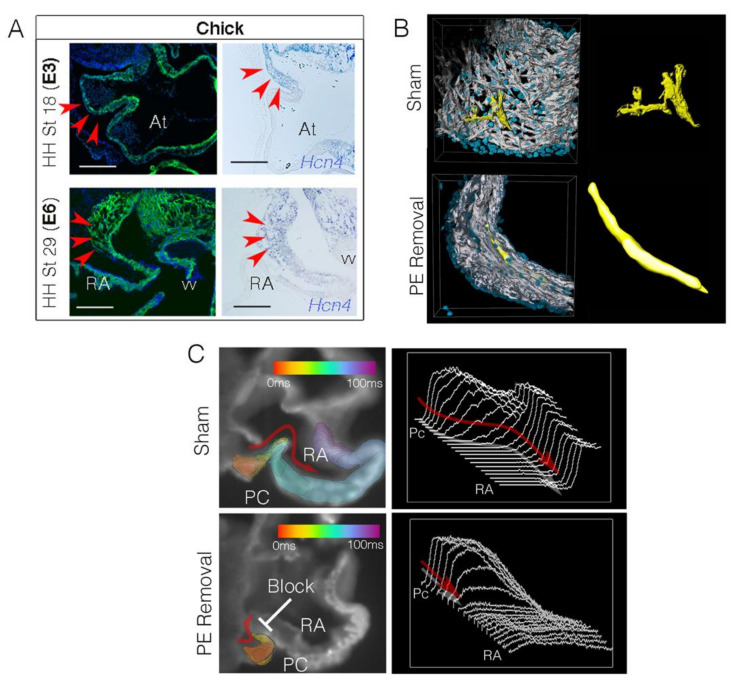
Figure 3.
Embryonic remodeling of the sinoatrial node. (A) Sections through the chick sinoatrial node at Hamburger Hamilton (HH) stage 18 [121] equivalent to embryonic day 3 (E3) and HH stage 29 (E6). Sections are stained with muscle marker MF20 (green) and Dapi (blue). In situ hybridization for the pacemaker cell marker Hcn4 indicates the location of the sinoatrial node (red arrowheads) (reproduced from [105]). Scale bar–200 mm. (B) Volumetric reconstructions of the chick sinoatrial node at HH stage 29. Data are shown from a control embryo (Sham) and an embryo in which proepicardial cells have been blocked from entering the sinoatrial node. MF20−white, Dapi−blue. Groups of pacemaker cells have been reconstructed in yellow to demonstrate the change in morphology when proepicardial cells are prevented from entering the sinoatrial node. (C) Isochronal maps of impulse propagation through sections of the sinoatrial node/atrial junction. Following proepicardial removal, sinoatrial node conduction block occurs (reproduced with permission from [104]). PC-pacemaker cells, RA-right atrium.

