Figure 3.
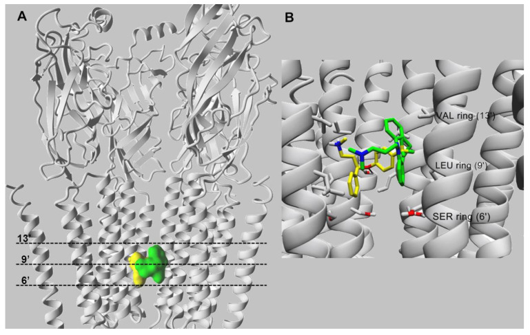
Molecular docking of fluoxetine (in yellow) and imipramine (in green), both in the protonated state, within the α4β2 nAChR ion channel (modified from [70]). (A) Side view of the overlapping binding sites for both ligands that interact with the middle portion of ion channel. (B) Imipramine (in green) and fluoxetine (in yellow) interact with the M2 transmembrane segments forming the lumen of the α4β2 nAChR ion channel. Both ligands interact mainly through van der Waals contacts with a domain formed between the valine (VAL) (position 13′) and serine (SER) (position 6′) rings. In addition, the black arrow indicates the hydrogen bond formed between the oxygen atom of fluoxetine and the hydroxyl group of α4-Ser251 (position 10′). For clarity, one β2 subunit is hidden. Residues involved in ligand binding are presented in stick mode (gray), whereas ligands are rendered either in the ball (A) or stick mode (B). All non-polar hydrogen atoms are hidden.

