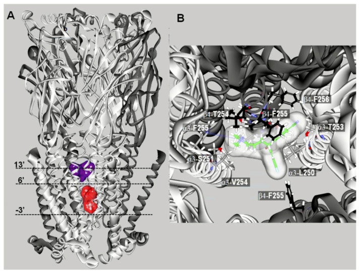
Figure 4.
Docking sites for S-(+)-citalopram (escitalopram) in the (α3)3(β4)2 nAChR model (modified from [68]). (A) Escitalopram docked at two luminal sites (surface model): a high-affinity site located closer to the extracellular ion channel’s mouth (blue) and a low-affinity site located closer to the cytoplasmic side (red). The α3 (white) and β4 (dark gray) subunits are represented as solid ribbons. Dotted lines indicate the positions of the Gly (position -3′), Ser (position 6′), and Val (position 13′) rings along the ion channel. (B) Detailed view of escitalopram at the high-affinity luminal site, showing the cation–π interaction with α3-F255 (position 14′) and the interaction with β4-T254 (position 12′).

