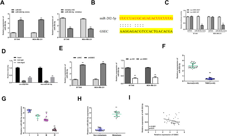
Figure 3.
GSEC served as a sponge of miR-202-5p. (A) BT-549 and MDA-MB-231 cells were transfected with miR-202-5p mimics/inhibitor or corresponding negative controls (miR-NC and inhibitor NC). The transfection efficiency was evaluated by qRT-PCR. (B) The putative targeting site between GSEC and miR-202-5p was predicted by Starbase v.2.0. (C) The luciferase reporter activity of Luc-GSEC-WT/MUT was detected by dual luciferase reporter assay. (D) The enrichment of GSEC and miR-202-5p was determined by RIP assay. (E) BT-549 and MDA-MB-231 cells were transfected with sh-GSEC, pc-GSEC (GSEC overexpression), or negative controls (sh-NC and pc-NC). The expression of miR-202-5p was evaluated by qRT-PCR. (F) The expression of miR-202-5p in TNBC tissues and adjacent normal tissues was evaluated by qRT-PCR (n = 45). (G) The expression of miR-202-5p in TNBC tissues of patients at Stage I (n = 11), Stage II (n = 9), Stage III (n = 12) and Stage IV (n = 13) was evaluated by qRT-PCR. (H) The expression of miR-202-5p in TNBC tissues of patients with lymph-node metastasis (n = 15) and non-metastasis (n = 30) was evaluated by qRT-PCR. (I) The correlation between GSEC and miR-202-5p in TNBC tissues was evaluated by Pearson’s correlation analysis (n = 45). ** p < 0.01.

