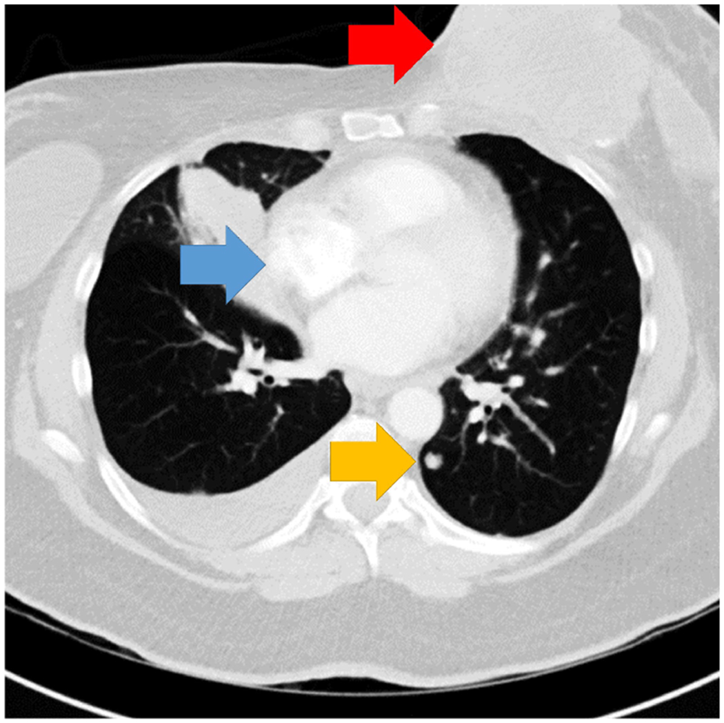
Figure 1.

Single-slice of the chest CT showing the abnormalities. Arrows indicate the location of the breast mass (red arrow), lymphadenopathy (blue arrow), and a lung nodule (yellow arrow). Arrows not present in experimental display.

Single-slice of the chest CT showing the abnormalities. Arrows indicate the location of the breast mass (red arrow), lymphadenopathy (blue arrow), and a lung nodule (yellow arrow). Arrows not present in experimental display.