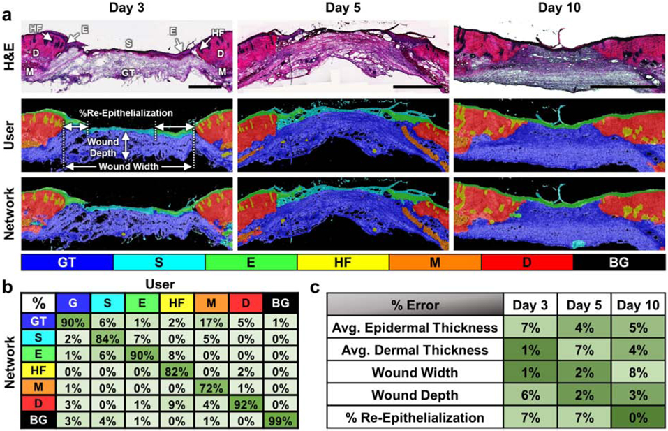
Figure 2.

Automated network segmentation and quantification of whole wound sections. (a) Representative H&E stained sections of skin wound tissue from days 3, 5 and 10 post-wounding (top row) with segmentation results from manual user tracing (middle row) and the CNN (bottom row) demonstrate the network’s ability to accurately segment full-thickness wounds. (b) The network demonstrated good accuracy across different wound regions and had an overall accuracy of 94.06%. (c) Automated measurements using the wound segmentation results revealed only small errors between the network and user-defined ground truth results. All scale bars are 500 μm.
