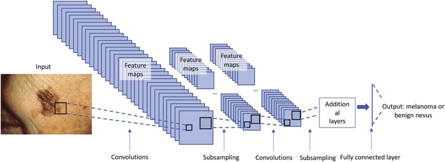
Figure 1:

During training, labeled image data (in this case skin lesions) is inputted to the neural network. Images undergo feature extraction (i.e. convolutions) and subsampling (i.e. pooling) steps in order for the network to learn the image features. As the network learns the image features, it adjusts weights to these features in order to optimize the correct classification of the input images. In this example, the output is whether an image represents a benign nevus or a melanoma.
