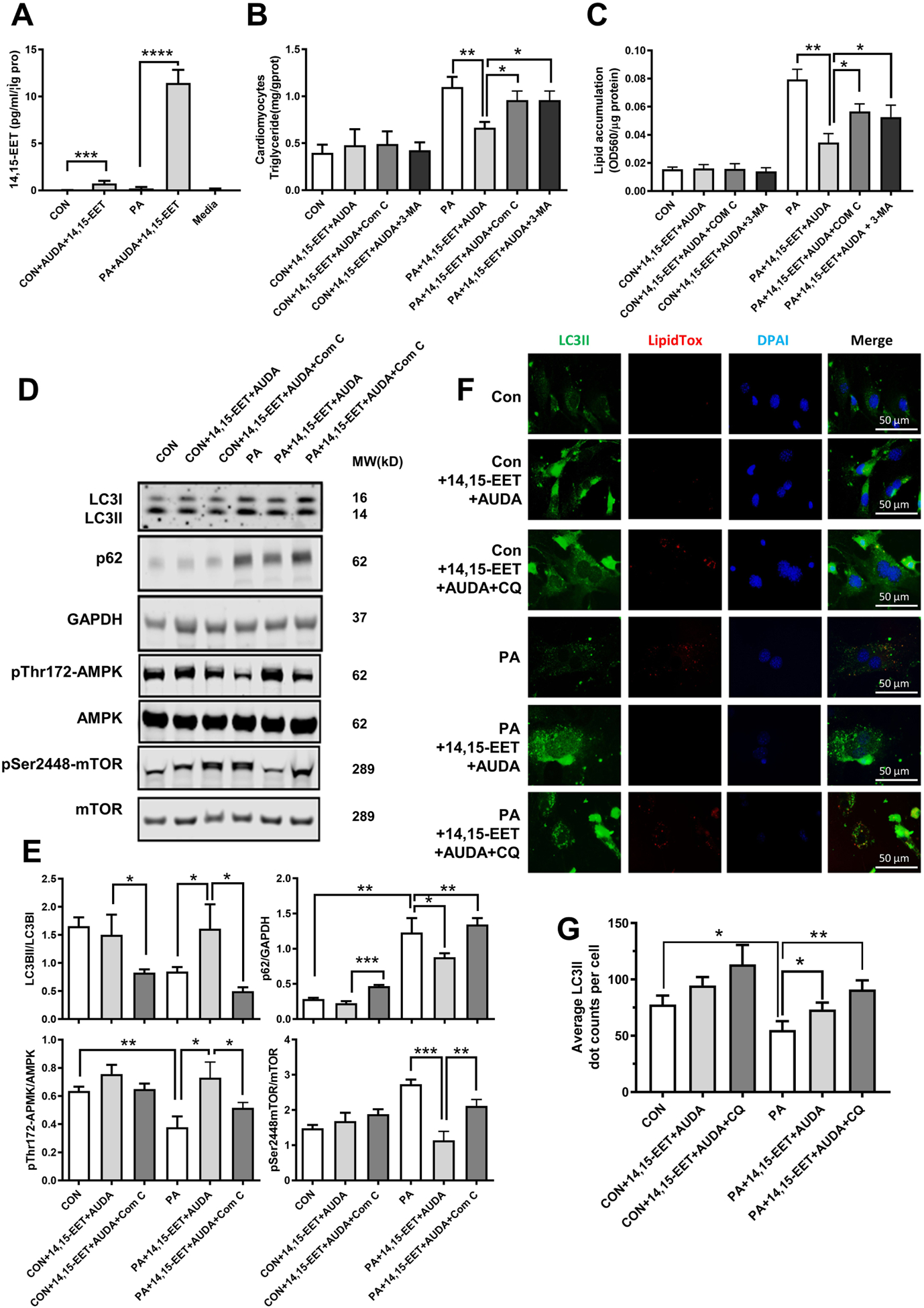
Fig. 5. Inhibition of AMPK or autophagy blocked the amelioration of lipid accumulation conferred by sEH substrate EET plus sEH inhibitor treatments in primary neonatal cardiomyocytes.

(A) Measurements of 14,15-EET in cardiomyocytes by liquid chromatography-mass spectrometry. (B) Triglyceride content of primary neonatal mouse cardiomyocytes from WT mice incubated with PA, 14,15-EET+AUDA, Compound C (Com C) or 3-MA. (C) Quantification of lipid accumulation with Oil Red O staining normalized to protein content (OD560/ μg protein). (D) Representative western blots of AMPK-mTOR-regulated autophagy pathway under Com C treatment. (E) Protein level quantifications of LC3II to LC3I, p62 to GAPDH, phosphorylated AMPK to total AMPK, and phosphorylated mTOR to total mTOR ratios. (F) Immunofluorescence images of LC3II and lipidTox. Scale bars indicate 50 μm. (G) Quantification of LC3II. *P<0.05, **P<0.01, ***P<0.005, ****P<0.001. n=3 per group. PA, palmitic acid; AUDA, sEH inhibitor; Com C, AMPK inhibitor; 3-MA, autophagy inhibitor; CQ, chloroquine; CON, no PA control; DAPI, 4’,6-diamidino-2-phenylindole; lipidTox, dye for lipid staining.
