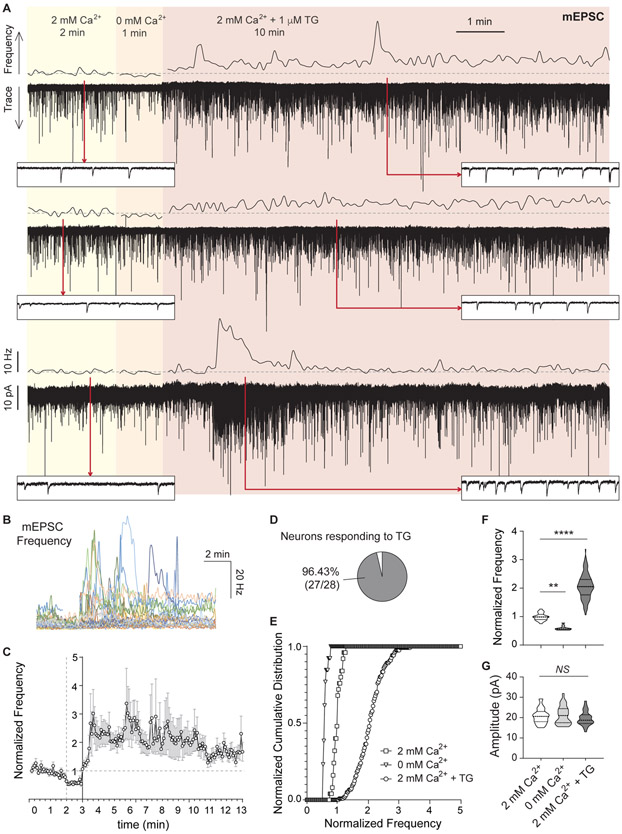
Figure 1. Ca2+ depletion from the ER augments spontaneous neurotransmission at excitatory synapses.
A. Three example traces (bottom of each pair) and 5 s moving averages of frequency (top of each pair) of mEPSC recorded from pyramidal hippocampal neurons. The same cell was continuously recorded while sequentially perfusing three different external solutions containing: 2 mM Ca2+ (2 min), 0 mM Ca2+ (1 min) and 2 mM Ca2+ with 1 μM TG (10 min). Dash line: average mEPSC frequency value of baseline (2 mM Ca2+). Insets show a 1 s window at the time points marked by red arrows.
B. 5 s moving averages of mEPSC frequency for all the experiments performed.
C. Time course of average ± SEM of normalized to baseline mEPSC frequency.
D. Percentage of patched neurons showing a positive increase in mEPSC frequency upon TG perfusion. For each recording, a positive effect was defined as: (Norm.freqTG-SDTG) > (Norm, freqbaseline + SDbaseline)
E-F. Cumulative histogram and violin plots of normalized mEPSC frequency.
G. Violin plot of mEPSC amplitude.

