
Fig. 3. Regions with significant associations between regional cortical thickness and latent trauma.
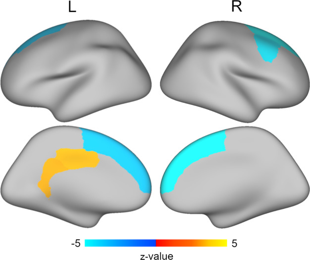
Structural equation modeling that controlled for age, sex, race/ethnicity, scanner model, and average cortical thickness revealed that greater latent trauma scores were associated with thinner cortices in bilateral superior frontal gyri and right caudal middle frontal gyrus (blue) and thicker cortices in left isthmus cingulate and posterior cingulate (yellow). Multiple comparisons were accounted for using the false discovery rate (q < 0.05).