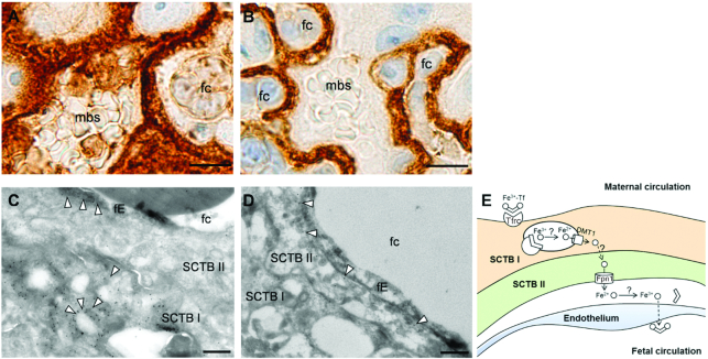
FIGURE 1.
Cellular localization of Tfrc and Fpn1 in the mouse placenta. IHC staining showing Tfrc expression in SCTB I (A) and Fpn1 expression in SCTB II (B) of E15.5 mouse placenta. Scale bar, 10 μm. High magnification immunogold EM images showing Tfrc on membranes of intracellular vesicles in SCTB and to a lesser extent on fetal endothelium (arrows) (C) and Fpn1 along the basal membrane of SCTB II (arrows) (D). Scale bar, 0.5 μm. (E) Updated mechanism of placental iron transport in the mouse placenta. DMT1, divalent metal transporter 1; E, embryonic day; EM, electron microscopy; fc, fetal capillary; fE, fetal endothelium; Fpn1, ferroportin 1; IHC, immunohistochemistry; mbs, maternal blood space; SCTB, syncytiotrophoblast; Tfrc, transferrin receptor.

