FIGURE 2.

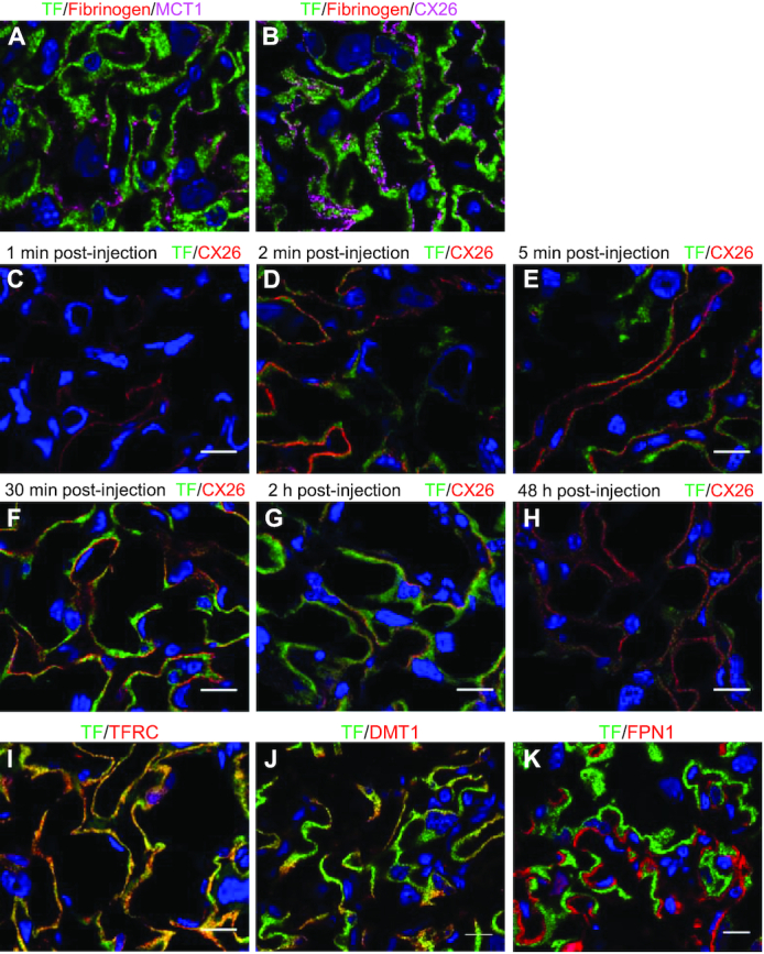
Placental transfer of maternally injected Tf. (A) In placentas collected 30 min postinjection, Tf (green) was fully endocytosed into SCTB I with no overlap with the apical membrane protein Mct1 (magenta) and (B) did not cross the border between SCTB I and SCTB II marked by the gap junction protein Cx26 (magenta). The negative endocytosis control injected simultaneously as Tf, fibrinogen (red), did not appear in SCTB I. (C–H) Accumulation and disappearance of injected Tf (green) in the mouse placenta within 48 h postinjection. Scale bar, 15 μm. Immunofluorescent staining of Tfrc (I), Dmt1 (J), and Fpn1 (K) in relation to Tf (green) in placentas 30 min post–Tf injection. Scale bar, 15 μm. Cx26/CX26, connexin 26; Dmt1/DMT1, divalent metal transporter 1; Fpn1/FPN1, ferroportin 1; Mct1/MCT1, monocarboxylate transporter 1; SCTB, syncytiotrophoblast; Tf/TF, transferrin; Tfrc/TFRC, transferrin receptor.
