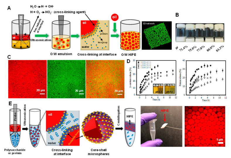
Figure 2.
(A) Schematic illustration of the preparation of HIPEs by ultrasonication. (B) Photographs of emulsions prepared by ultrasonication at different volume fractions of oil (ϕ). (C) Confocal laser scanning microscopy (CLSM) images of HIPEs, showing the oil phase stained red (left), the protein-rich aqueous phase stained green (middle), and the merged images (right). (D) Release profiles of β-carotene and bovine serum albumin (BSA) from HIPEs during incubation under simulated physiological solutions at pH 1.2 and pH 7.4. The inset shows the visual appearance of the HIPEs taken after 6 and 12 h incubation at pH 1.2 and 7.4, respectively. HIPEs prepared by ultrasonication and homogenization are labeled as U-HIPEs and H-HIPEs. Reprinted with permission from [67]. Copyright (2018), American Chemistry Society. (E) Schematic illustrating the preparation of polysaccharide- and protein-based HIPEs through successive ultrasonication and centrifugation, right corner is the CLSM image of HIPEs stabilized by chitosan and pectin. Reprinted with permission from [70]. Copyright (2018), Royal Society of Chemistry.

